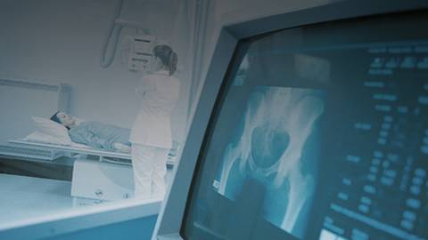
ULTRA-PORTABLE
ULTRASOUND IN THE CRITICAL CARE UNIT
You are listening to ReachMD XM160, The Channel for
Medical Professionals. Welcome to Advances in Medical Imaging, a program
discussing the latest innovations in clinical radiology and imaging
technologies. Your host is Dr. Jason Birnholz, Director of Diagnostic
Ultrasound Consultants in Oak Brook, Illinois.
Making critical care medicine faster and more efficient.
You are listening to ReachMD, The Channel for Medical Professionals. Welcome
to Advances in Medical Imaging. I am Dr. Dr. Jason Birnholz, your host and
with me today is Dr. Eyal Herzog, who is an Assistant Professor of Clinical
Medicine at the Columbia University College of Physicians and Surgeons where he
is working on a streak of 4 Consecutive Teacher of the Year Award and he is
Director of Cardiac Care at the St. Luke's-Roosevelt Hospital in New York
City. Today, we are discussing pocket ultrasound in critical care.
DR. JASON BIRNHOLZ:
Hello, Dr. Herzog, thank you for joining us. I used the
term pocket ultrasound as the way of illustrating that the new ultra-portable
ultrasound units are quite small. How do these units compare in performance
with the larger devices?
DR. EYAL HERZOG:
Yes, they are small and we can actually put them in our
pockets. We did study. We did compare the imaging quality of the small, we
actually call them echoscopes, this is our term for them, compared to our
high-end echo machines and surprisingly at least for the 2-dimension imaging
that we obtain in critical care setting, we were very happy to find that these
imagings are excellent and equivalent to the high-end machines.
DR. JASON BIRNHOLZ:
Oh, good, once you have a cardiologist or let's say a
hospitalist with critical care patients or an internist that has mostly cardiac
patients and they are not currently using ultrasound, would they be comfortable
with one of these small units or should they start with something else?
DR. EYAL HERZOG:
I mean, they have to be trained first. At least, the study
we had performed and the performer or the operator of the device well echo
technicians or cardiology fellows or attendings who are trained to perform
ultrasound. So, it's not yet applicable to all physicians because they have to
be first trained to perform and interpret an echo imaging.
DR. JASON BIRNHOLZ:
That's true of any procedure essentially. Well, I mean the
units are a bit fragile. They are expensive. Now, are they meant to be
carried around mainly by a physician or in your unit, you leave them in a
drawer and have anybody on the unit use it who of course is competent to use
it?
DR. EYAL HERZOG:
Yeah, if you have a competent user, well we can leave them
in the unit or next to the unit and when the patient presents, you know, they
can just pick it up and use it or if the on-call physician is in the unit, he
can carry it the same way he carries his stethoscope.
DR. JASON BIRNHOLZ:
Oh, well do you carry one around?
DR. EYAL HERZOG:
I do not carry one around because at this point, this is
only a research tool for us and we do not use it as yet. As a clinical tool to
you, we are actually showing that it's the placing the tool.
DR. JASON BIRNHOLZ:
Oh, I was hoping you are going to say – Oh yes, I carry it
around and I use it on morning rounds to check people instead of using my
stethoscope.
DR. EYAL HERZOG:
No, this is we are not there yet. We are far away from
replacing the stethoscope.
DR. JASON BIRNHOLZ:
Suppose you are asked to see a patient, he is brought into
the emergency room, he is diaphoretic, and he has chest pain. Let's say, you
know, he is alert and stable with don't know wild factors, you are there to see
him, you have this portable unit, would you kind of step through what you think
you might see or want to look at?
DR. EYAL HERZOG:
We have to be very careful here because at this point, we
just compare the quality of imaging to test this is a part of this is recent
device tool to use. So, at this point, as of today, we are actually not using
it in our clinical scenarios when we are actually facing patients. However, a
few years down the road, a few months down the road, when this matures and the
technology improved, I do believe that there is a room to carry it, you know, a
cardiologist should carry this kind of transducer, which is not heavy and you
can actually have it in your pocket. It will actually help you to diagnose the
patient. It is not going, I mean you are still going to examine the patient.
DR. JASON BIRNHOLZ:
Yeah, sure, I mean I just want to short circuit that and in
the sense if you want to think back to using a conventional echocardiography unit
and that you wheeled up to the patient, it really should be the same since you
said the image quality is similar.
DR. EYAL HERZOG:
Again, it's the question of the maturity of the device. The
device we are testing, yet they do not have a color flow yet, so if you don't
have a color flow, you still cannot make a diagnosis of leaking valve or mitral
regurgitation or aortic regurgitation, etc., which are critical in critically
ill patient. So, you still need to examine the patient for murmurs. However,
I do believe that within the next 6 or 12 months, the manufacture will have these
color imagings with the small echo units. At this point, I do believe that
actually if you are trained to do it, it can replace the full-sized echo
machine in critical care setting where you do not need to get the entire
information that you obtain with a full echo.
DR. JASON BIRNHOLZ:
For a lot of people, who are not doing ultrasound and order
studies, but are not quite aware of what the physician gets when looking
directly and you know what to look for, so if you want to step back to a few
years ago and you have a 2-dimensional echocardiography unit without color flow
for sample and you are faced with a patient that has chest pain and you have
examined him, you know, just in terms of the ultrasound, you are putting the
probe on, looking, what do you tend to look for, what do you think of, what
might you say?
DR. EYAL HERZOG:
What I have done over the years, I have developed different
pathways for management of acute coronary syndrome, chest pain, and heart
failure. One thing is for sure, if someone comes in for acute myocardial
infarction or to be proper size, the ST elevation myocardial infarction, we do
not want to waste any time for imaging anyway. We base our clinical decision
based on the electrocardiogram and based on the patient’s symptom, if there is
clear ST elevation in the EKG, the patient goes to the cath lab for pulmonary
angioplasty. Imaging delays care in this patient. So, imaging comes to the
management of the acute coronary syndrome and heart failure patient only when
you really do have the time to obtain the imaging and not to delay care. At
that point, if you do not want to wheel the big echo machine, you don't have it
available there, then this small machine can help you. For example, cases
that's related to critical care can be a pericardial effusion, assess of wall
motion analysis or wall motion abnormality in the setting of a normal EKG for
example, when you are not certain what's the next step in management of
patient, but it has to be very clear. If you really have this ST elevation
myocardial infarction, you do not, you do not perform an echo, you are just
going to delay the care.
DR. JASON BIRNHOLZ:
If you are just joining us, you are listening to Advances in
Medical Imaging on ReachMD, The Channel for Medical Professionals. I am Dr.
Jason Birnholz and I am speaking with Dr. Eyal Herzog. We are discussing
pocket ultrasound in critical care.
DR. EYAL HERZOG:
So, this is actually a very important question. When
someone presents with acute heart failure for example, then you may want to
diagnose if we are taking about LV systolic dysfunction where there is decrease
in left ventricular function and motion. In this situation, this small tool
can help you to identify the wall motion, it will identify that the patient’s
ejection fraction is reduced or severely reduced and this can help you, for
example, in diagnosing this group of patient for patient with hypertrophic
cardiomyopathy who come presenting with the same symptoms of acute heart
failure. However, they have very thick walls with hyperdynamic function of the
heart and the treatment will be completely opposite. In the first group of
patient, you want to give a medication that will improve the function of the
heart while in the second group of patient, you want to give a medication,
which actually will reduce the function of the heart, for example
beta-blocker. So, this tool will be great to identify acute heart failure, to
identify if it is systolic dysfunction or hypertrophic cardiomyopathy for
example. So, this is great at it in the situation.
DR. JASON BIRNHOLZ:
What about if you have the situation of an ambulance, where
you have the transit time and you've stabilized the patient, is there a role
there for these units?
DR. EYAL HERZOG:
If the performance of the procedure will not delay care,
then it can be helpful. For example, evaluation of pericardial effusion and
tamponade. You may want or may not want to use specific therapy if you do have
pericardial effusion in the case of myocardial infarction, for example. It is
great to evaluate mechanical complication of myocardial infarction. Again, we
are still waiting for the color imaging to be added to this modality; however,
if you have someone with a new murmur when you examine the patient and you want
to evaluate why they have this new murmur, you can exclude mechanical
complication for example, mitral regurgitation, free wall rupture with
pericardial effusion, ventriculoseptal defect, right ventricular function.
These are all mechanical complications that may, if you have the time in the
ambulance, can lead to better treatment for the patient.
DR. JASON BIRNHOLZ:
Now, are you just looking at the wall movement patterns or
are you documenting these in some way or are you doing any?
DR. EYAL HERZOG:
When we use it, we still use it as a research tool. So,
what we do in all our patients is to get a full study by the high-end echo machine
and we get the best that the small echo machine can provide, so we can measure
wall thickness, we can grade wall motion, we can grade and measure pericardial
effusion if it exists. We can evaluate valve motion; however, we cannot yet
evaluate valve regurgitation because we don't have the color flow and these are
the basic things we do at this point.
DR. JASON BIRNHOLZ:
When you are using these units, I mean, experimental or
otherwise, are their findings documented in some way or is this very much like
fluoroscopy where you look and you say "Aha, I see something" and
then you write it down?
DR. EYAL HERZOG:
You type it, it's a small computer, you get a video screen
that you can type and you save it. It's all digital. You download to our
computer and you can analyze it later or same time of obtaining the imaging,
but you can view it later on. It's all digital imaging.
DR. JASON BIRNHOLZ:
Oh, so you can determine ejection fractions and do more
quantitative things.
DR. EYAL HERZOG:
Sure, you can bring it later to your computer, to your
desktop, download the imaging and measure things that you want to measure.
DR. JASON BIRNHOLZ:
What about using this in conjunction with stress testing.
DR. EYAL HERZOG:
This is too early. I mean, we do have small echo machines,
not what we call the echoscope or the pocket echos, but small hand echo
machine, some of them do have the feature of performing stress testing, but I
don't think this is where this tool is going in the right direction because
when you bring someone for a stress test, in anyway you want the best quality,
the best imaging and you are going to use the high-end machine. We do a lot of
stress testing in our institute with echo imaging.
DR. JASON BIRNHOLZ:
Well, then let me turn that around the other way. Since you
are in a critical care unit, I am sure you have every possible kind of
equipment anybody can ever want on wheels to be brought anywhere instantly or
this I would imagine that why are you thinking of using this at all?
DR. EYAL HERZOG:
Okay, so let's go to the way how a real critical care unit
sick patient looks like, there are probably like fixed drips coming through
the, you know, the veins of the patient’s ventilator, occupying space in the room
and you really do not have the feasibility to bring this large high-end echo
machine next to the patient in the right position and you know things are
changing every second. So, if you really want a tool that will examine the
patient with things happen, you have to be fast and things can happen in 10
minutes and we can re-image the patient in another 10 minutes when you round on
another patient. So, this you cannot just wheel and although, you know, heavy
machine from room to room and there is space even though in all our units in
our large room, but still in very sick patients, usually the room are busy with
a lot of equipment, you know.
DR. JASON BIRNHOLZ:
What are these things that you find from the small unit,
let's say you are using this all the time that your other monitoring things on
these critically ill patients are not showing you.
DR. EYAL HERZOG:
The key is timing in critical care, it's feasibility, you
have it, you can make a quick diagnosis, it can help you to save patients. We
wheel patient directly from our CCU or based on imaging to the OR for a case of
aortic dissections, tamponade, pericardial effusion, which led to tamponade
situation and this is likely.
DR. JASON BIRNHOLZ:
My thanks to Dr. Eyal Herzog, who has been our guest. We
have been discussing pocket ultrasound in critical care. Dr. Herzog, thank you
very much. I hope you will keep us updated on your work.
DR. EYAL HERZOG:
Okay, thank you very much.
DR. JASON BIRNHOLZ:
I am Dr. Jason Birnholz and you have been listening to
Advances in Medical Imaging on ReachMD, The Channel for Medical Professionals.
You have been listening to Advances in Medical Imaging.
For more details on this week's show or to download this segment, visit us at
reachmd.com. Thank you for listening.


Facebook Comments